Bertholletia
Placentation as seen in ovary longitudinal section of Bertholletia excelsa.

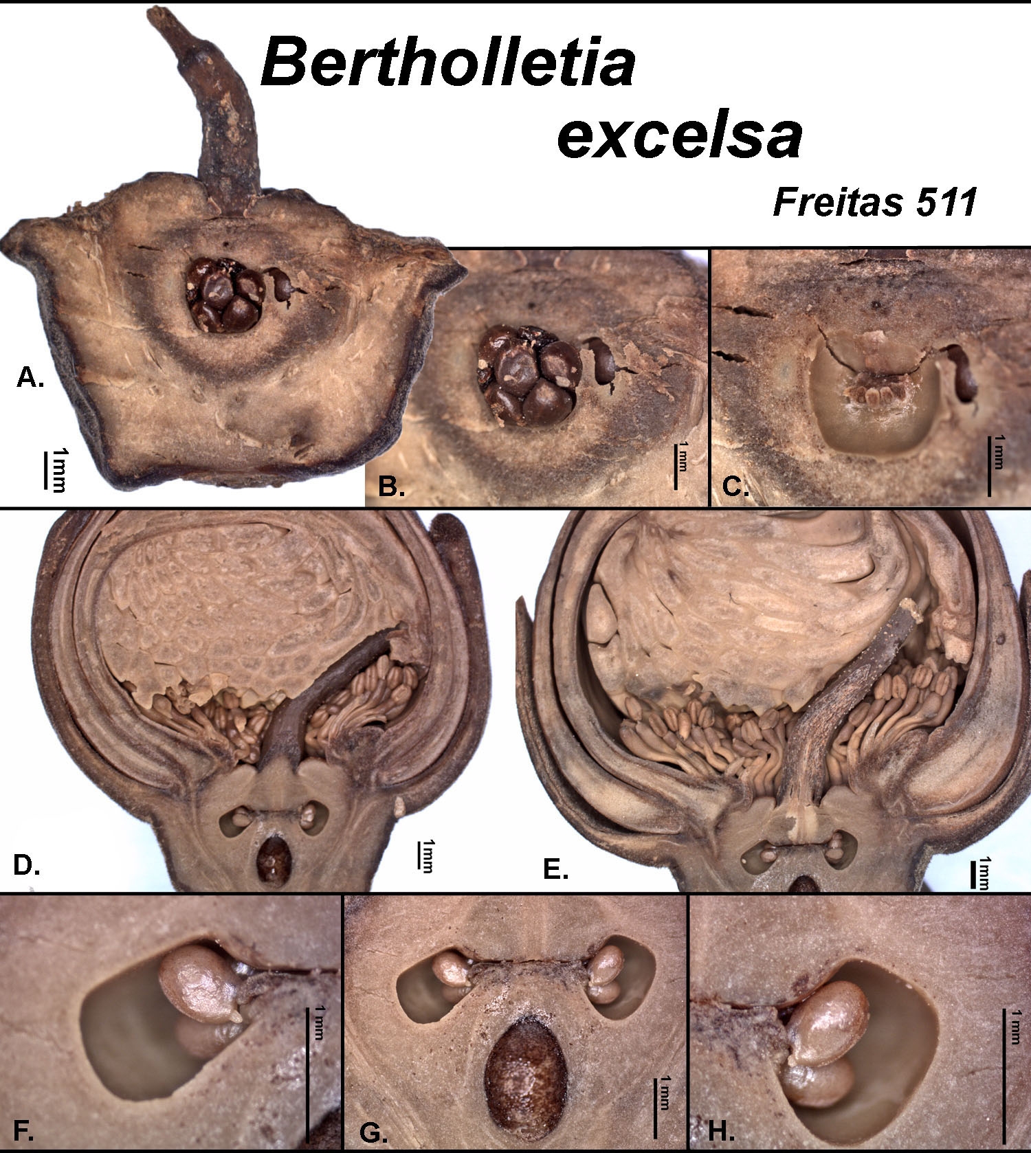
Placentation of Bertholletia excelsa as shown in longitudinal sections based on Freitas 511 from Amazonas, Brazil. A. Section through the wall of a locule showing the ovules. B. Close-up of A. C. Locule after the ovlues have been removed. D. Medial section of a bud showing the locules at the apex of the septum. E. Close-up of D. F. Close-up of a locule on the left side of section G. Note the extruded inner integument of the ovule. G. Section showing the two locules plus a chamber of unknown function. H. Close-up of the locule on the right side of section G.
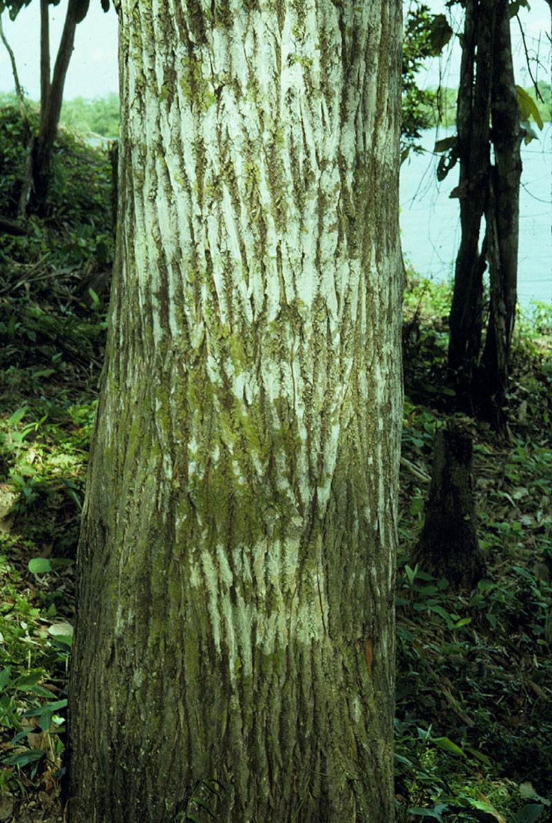
Fissured bark of a Brazil nut tree.
The fissured bark of a Brazil nut tree (Bertholletia excelsa) based on an unvouchered tree from the Rio Negro, Brazil.
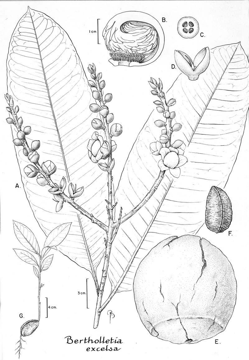
Botanical line drawing of Bertholletia excelsa. Drawing by B. Angell.
Fig. 45 from Fl. Neotrop. Monogr. 21(II). 1990. A. Habit, note how the petals are tightly pressed against the androecium. B. Medial longitudinal section of an androecium, note how the hood appendages are swept inwards but do not form a complete coil. C. Cross section of an ovary. D. Ovary and calyx, note the 2-parted calyx, sessile ovary, and long, geniculate style. E. Fruit, note that the opercular opening is smaller in diameter than the size of the seeds. F. Seed. G. Seedling.