Bertholletia excelsa (Image keywords)
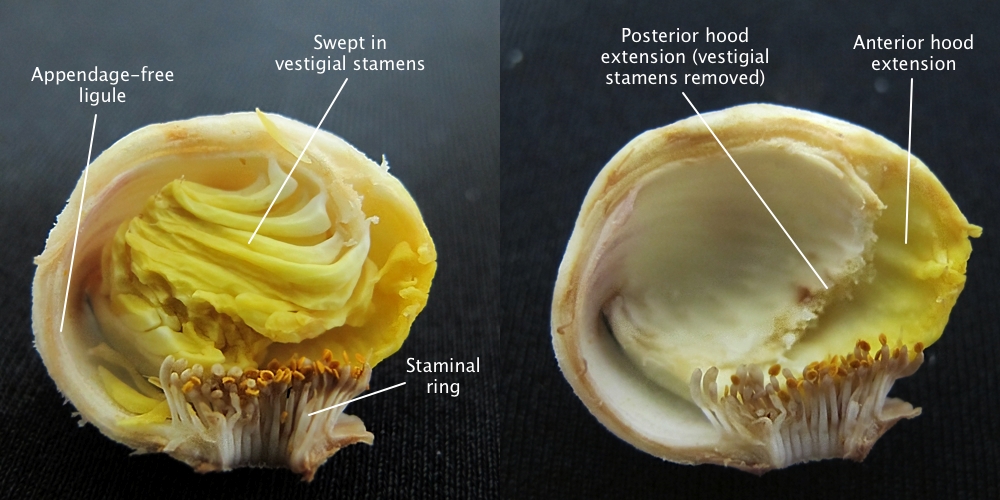
Medial sections of androecial hoods of Bertholletia excelsa.

Medial sections sections of an androecium of Bertholletia excelsa based on an unvouchered tree, Amazonas, Brasil. Left: section with the vestigial stamens in place to show their swept-in orientation and split into a posterior hood extension and an anterior hood extension. Note the clavate filaments of the outermost stamens (right) and the unidimensional filaments of the inner stamens. The distance from A to C is the ligule and from B to C is the androecial hood (including the vestigial stamens.
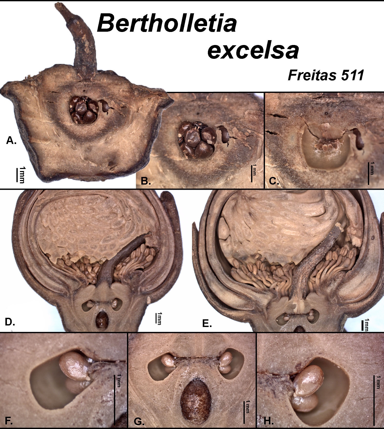
Placentation as seen in ovary longitudinal section of Bertholletia excelsa.
Placentation of Bertholletia excelsa as shown in longitudinal sections based on Freitas 511 from Amazonas, Brazil. A. Section through the wall of a locule showing the ovules. B. Close-up of A. C. Locule after the ovlues have been removed. D. Medial section of a bud showing the locules at the apex of the septum. E. Close-up of D. F. Close-up of a locule on the left side of section G. Note the extruded inner integument of the ovule. G. Section showing the two locules plus a chamber of unknown function. H. Close-up of the locule on the right side of section G.
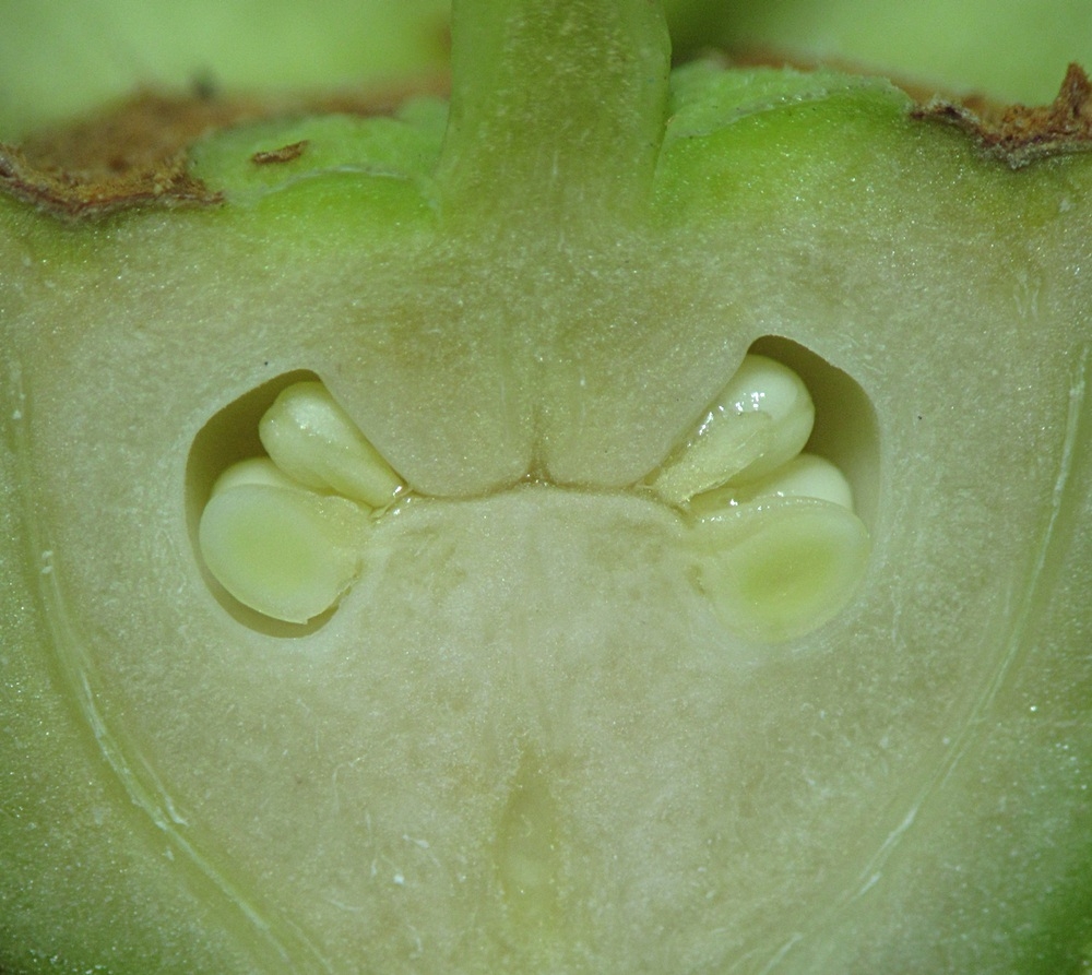
Placentation as seen in ovary longitudinal section of Bertholletia excelsa.
Placentation of Bertholletia excelsa as seen in a medial longitudinal section based on N. P. Smith 397 from the Bosque da Ciência, Instituto Nacional de Pesquisas da Amazônia (INPA), Manaus, Amazonas, Brazil.
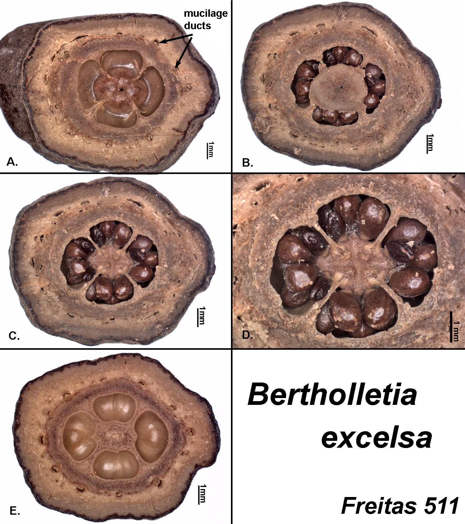
Placentation as seen in ovary cross-sections of Bertholletia excelsa.
Placentation of Bertholletia excelsa as shown in cross-section based on Freitas 511 from Amazonas, Brazil. A. Looking from the base toward the apex of the ovary. Note that ovules are attached above this point. B. Looking from the apex toward the base of the ovary. The round cap in the center was lifted without tearing it away from other tissue to reveal the view seen in the next image. C. View same as in B. Note this view shows the top of the placenta and the uppermost ovules attached to it. D. Close-up of C. Note the broadly triangular shape of the placentae which arise at the ends of the septa. E. An apical view of the very base of the locules, i.e., of an ovule-free area below the placentae. The locule number of four is relatively consistent in this species.
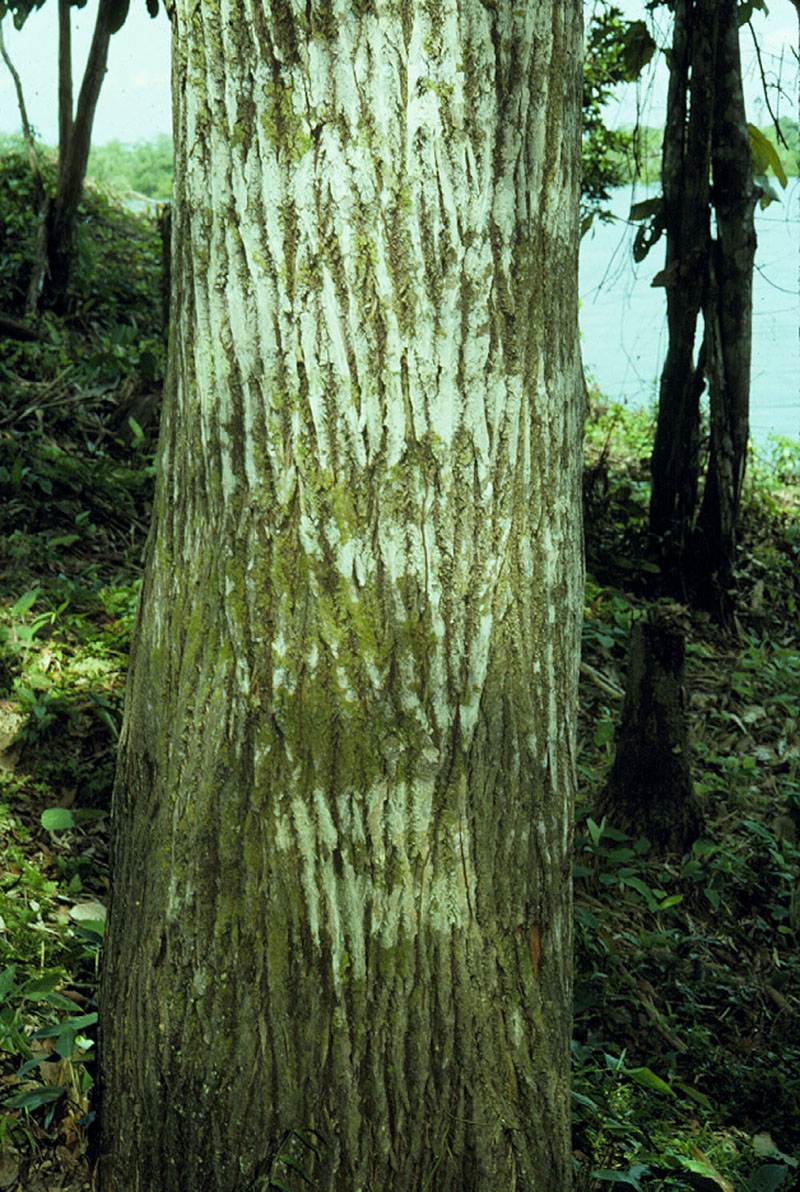
Fissured bark of a Brazil nut tree.
The fissured bark of a Brazil nut tree (Bertholletia excelsa) based on an unvouchered tree from the Rio Negro, Brazil.
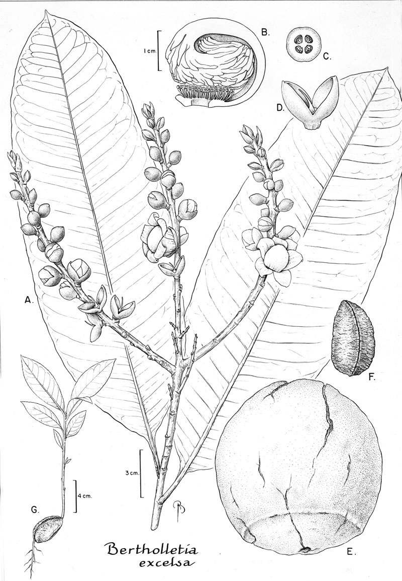
Botanical line drawing of Bertholletia excelsa. Drawing by B. Angell.
Fig. 45 from Fl. Neotrop. Monogr. 21(II). 1990. A. Habit, note how the petals are tightly pressed against the androecium. B. Medial longitudinal section of an androecium, note how the hood appendages are swept inwards but do not form a complete coil. C. Cross section of an ovary. D. Ovary and calyx, note the 2-parted calyx, sessile ovary, and long, geniculate style. E. Fruit, note that the opercular opening is smaller in diameter than the size of the seeds. F. Seed. G. Seedling.
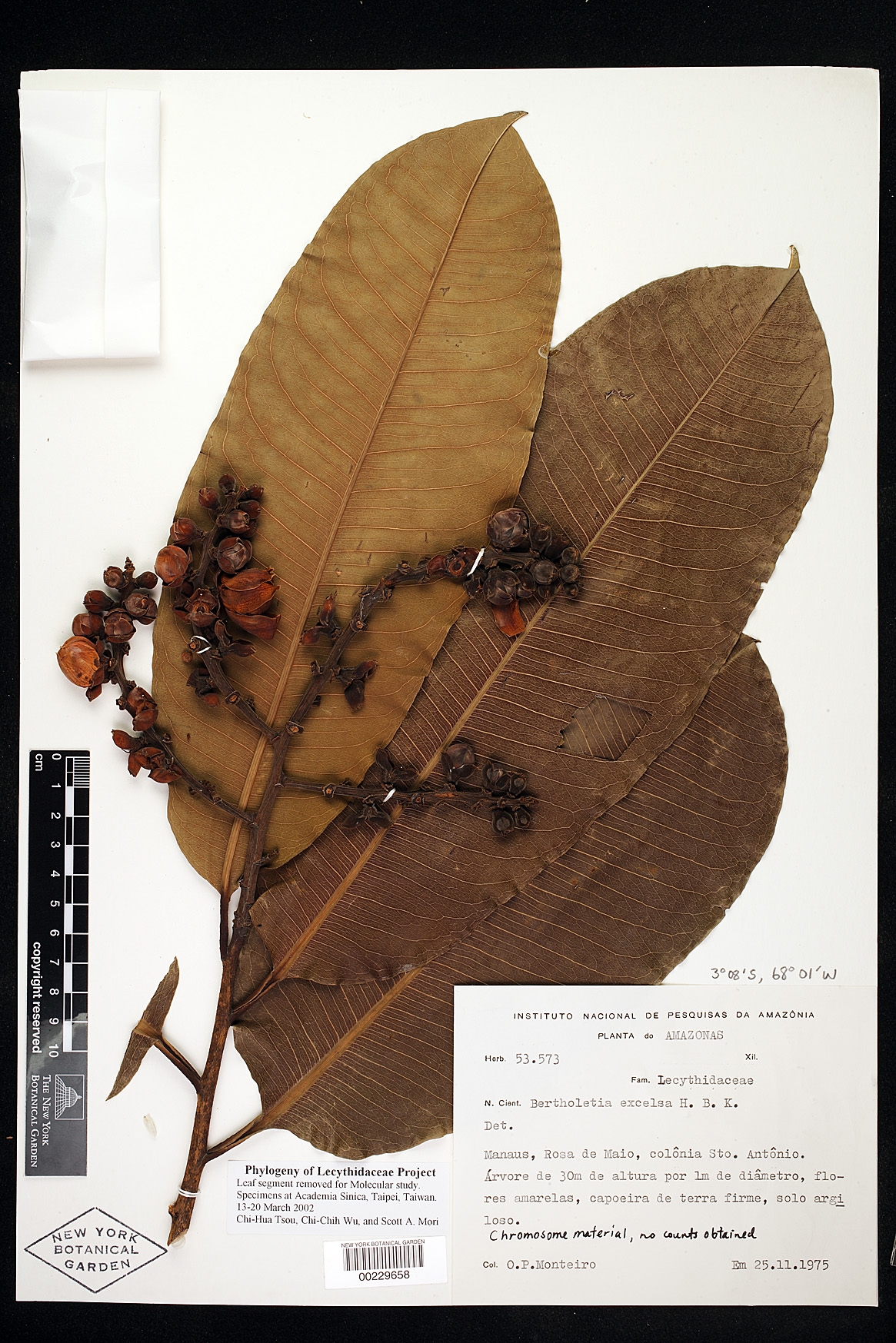
Herbarium sheet of Bertholletia excelsa
Herbarium sheet of Bertholleltia excelsa based on O. P. Monteiro INPA 53573 from the state of Amazonas, Brazil. Note the canaliculate petioles; the relatively large, oblong leaf blades; once-branched inflorescences; and buds with two-lobed calyces.
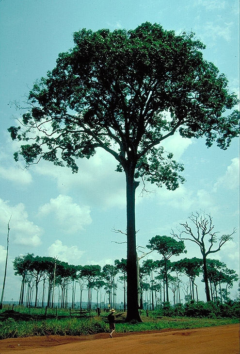
Remnant Brazil nut trees (Bertholletia excelsa)
Remnant Brazil nut trees (Bertholletia excelsa) based on unvouchered trees from Mato Grosso, Brazil. Deforestation is the major threat to the survival of this species.